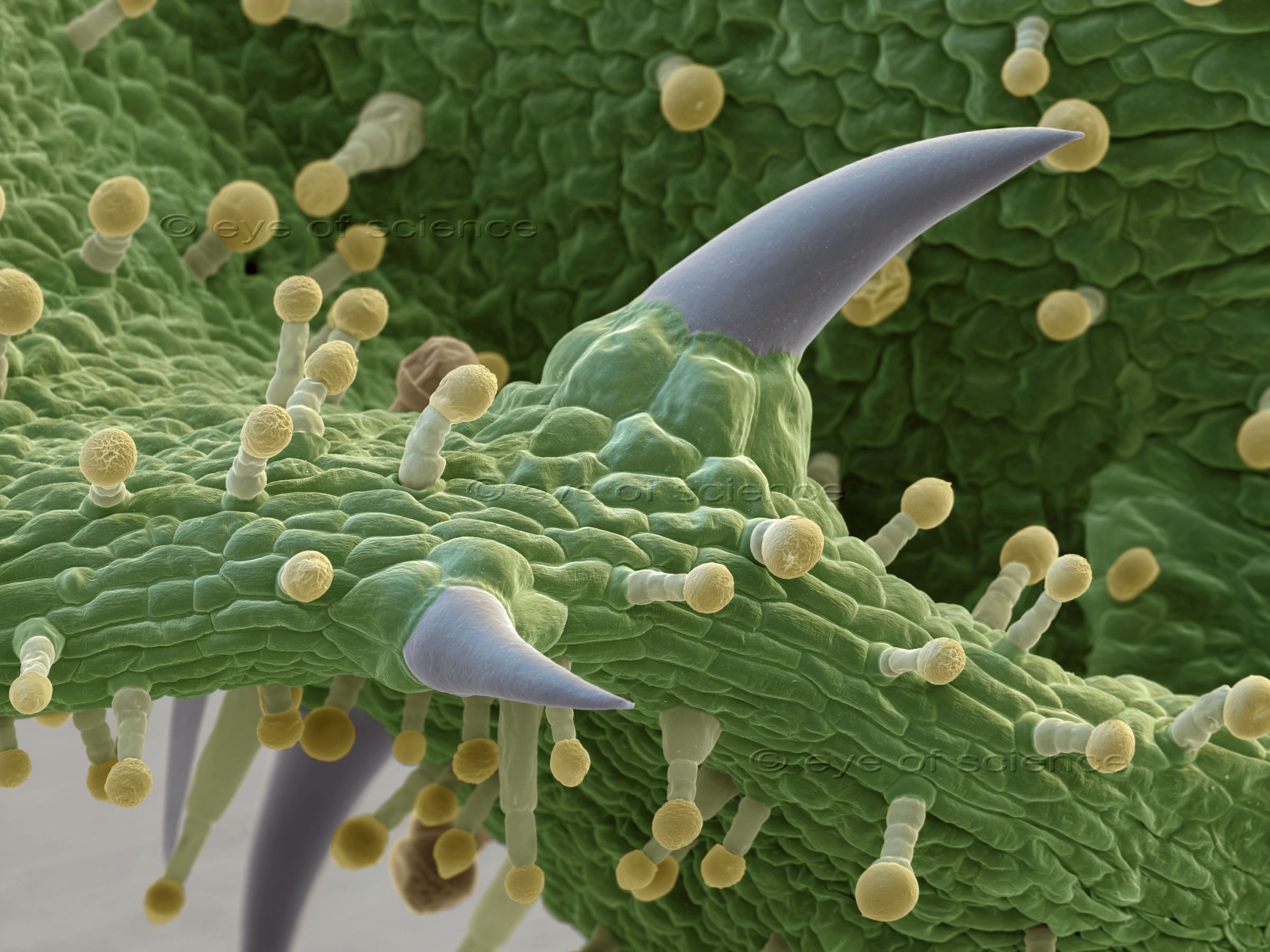
Low magnified SEM image of Chamomilla
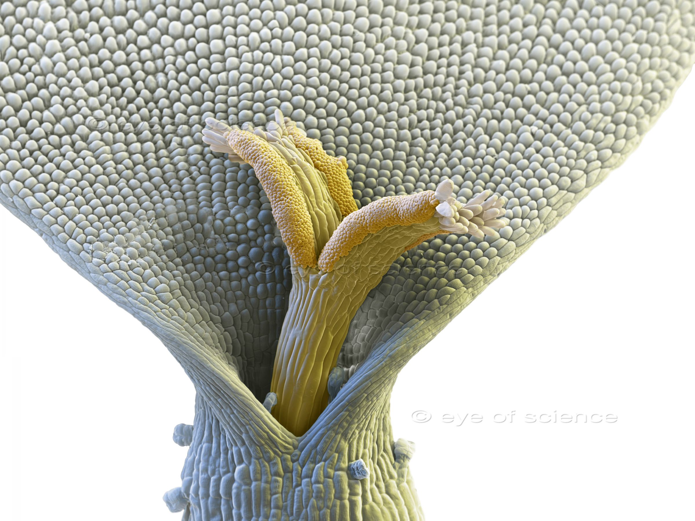
Single blossom of a Chamomile flower. Scanning Electron Microscope, 170:1 (when 15cms wide)
Scanning Electron Micrograph of Hamamelis
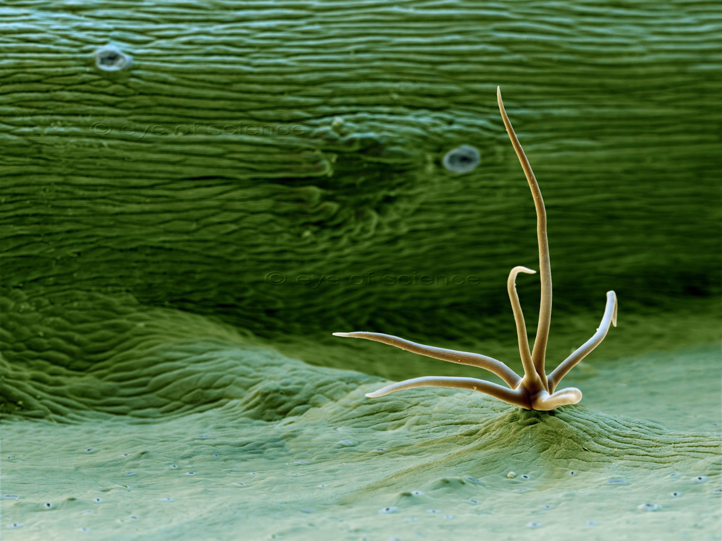
Trichome (leaf hair) on a leaf of Hamamelis. Scanning Electron Microscope, 170:1 (when 15cms wide)
SEM image of Fennel
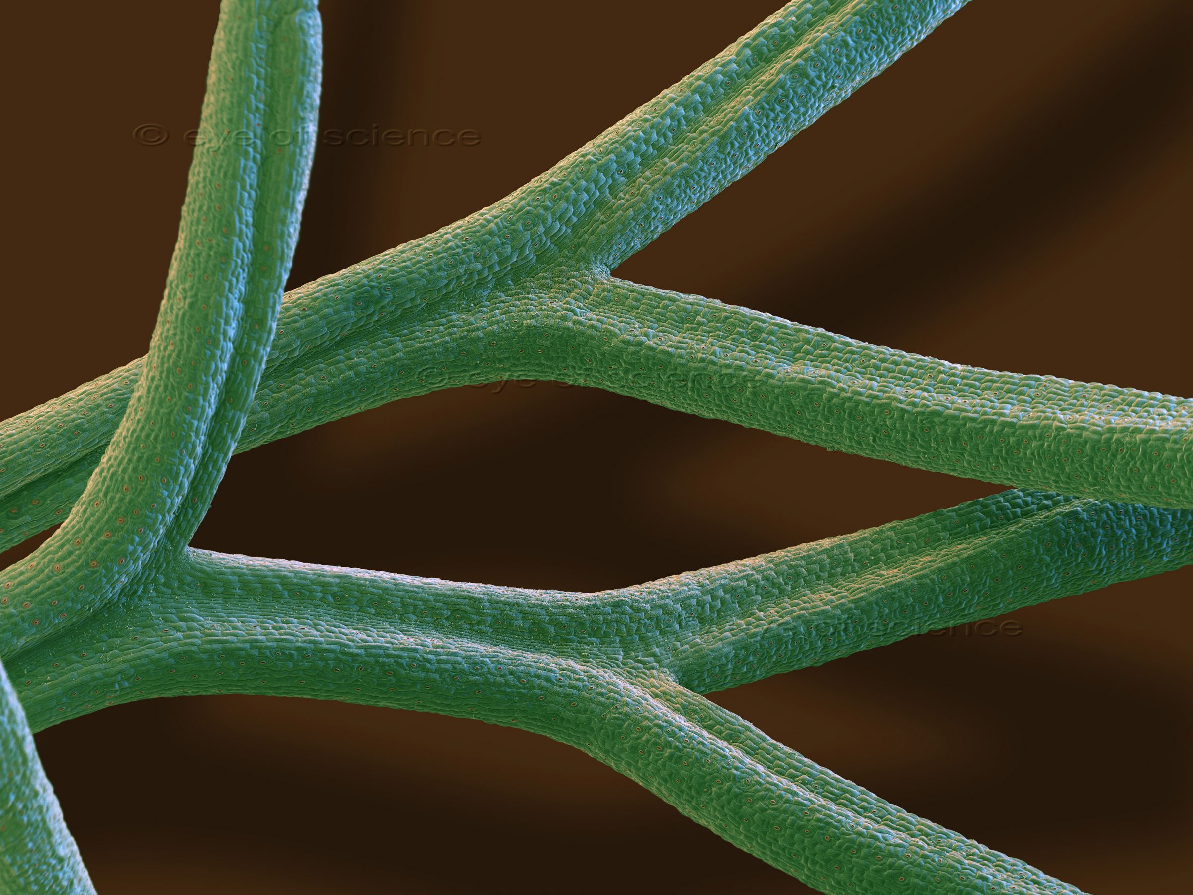
Fennel leaves with tiny stomata (brown spots) all over. Scanning Electron Microscope, 50:1 (when 15cms wide)
Fern seen with an Electron Microscope
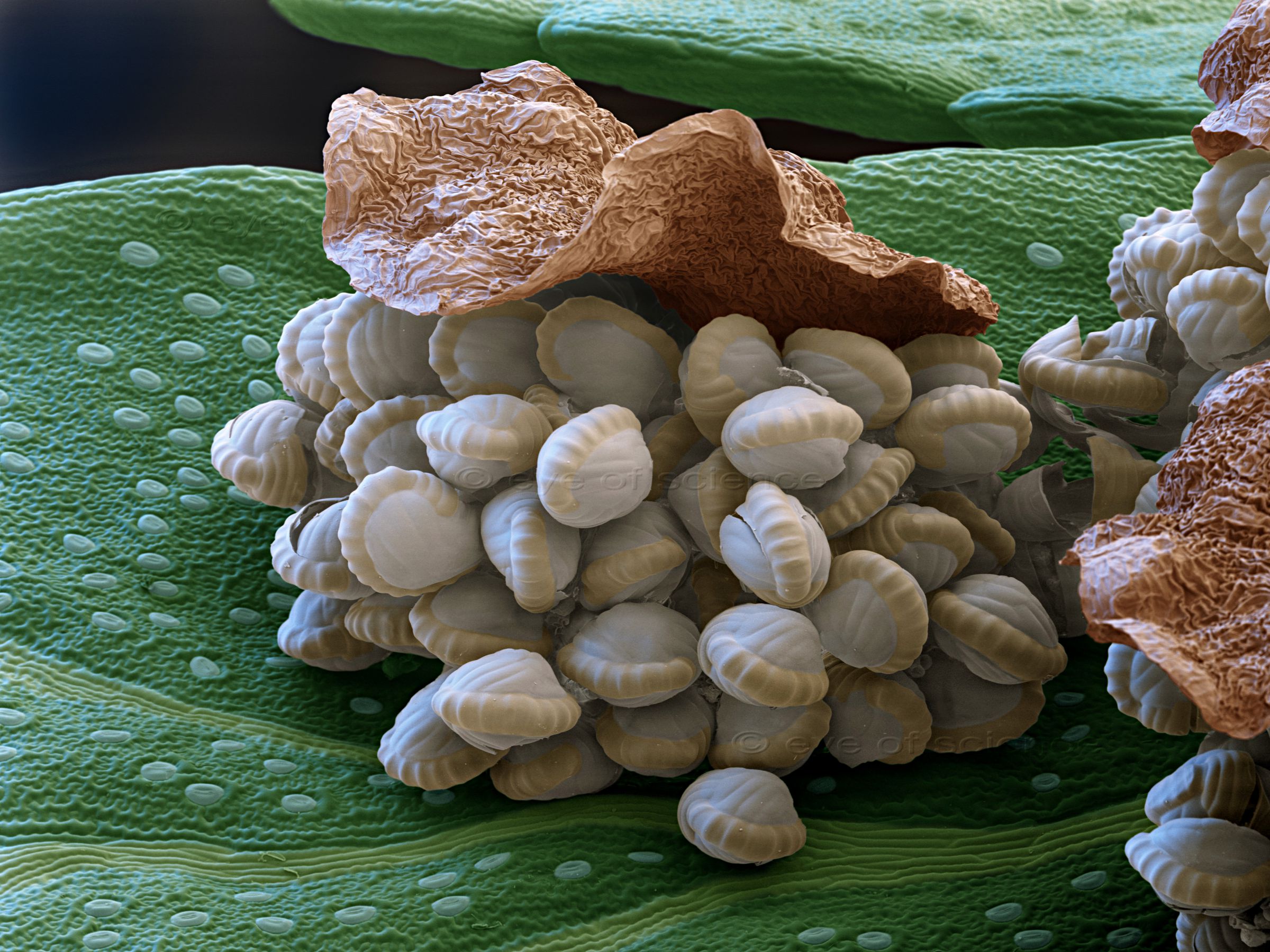
Open sorus of the male fern (Dryopteris filix-mas). The spore capsules that contain dozens of spores, are shown whitish here. Scanning Electron Microscope, 80:1 (when 15cms wide)
Micrograph of a Coffee Bean
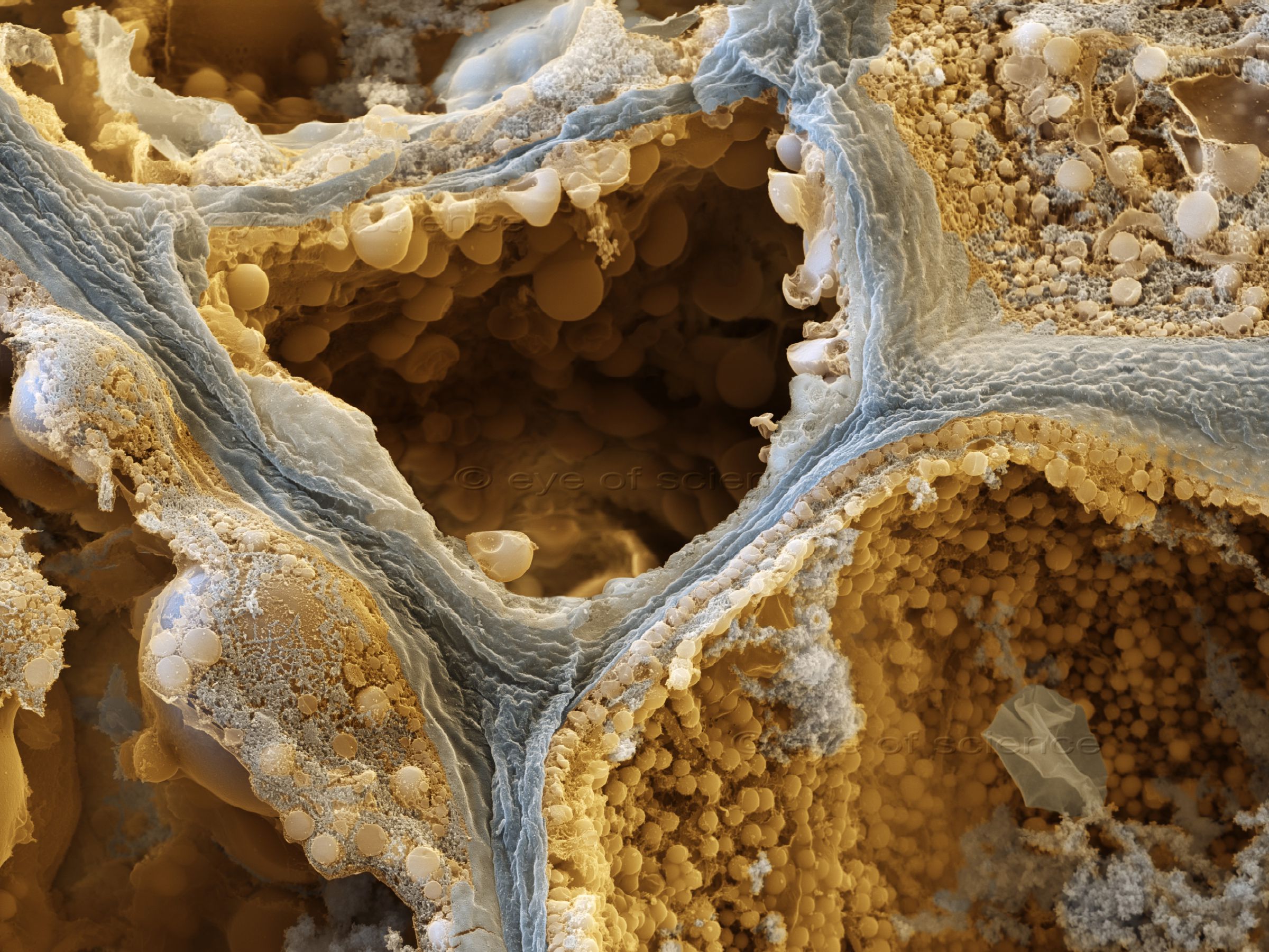
Freeze fraction of a Coffee bean showing the content of the cells. Scanning Electron Microscope, 5000:1 (when 15cms wide)
A Leaf Section shows Microscopic Details.
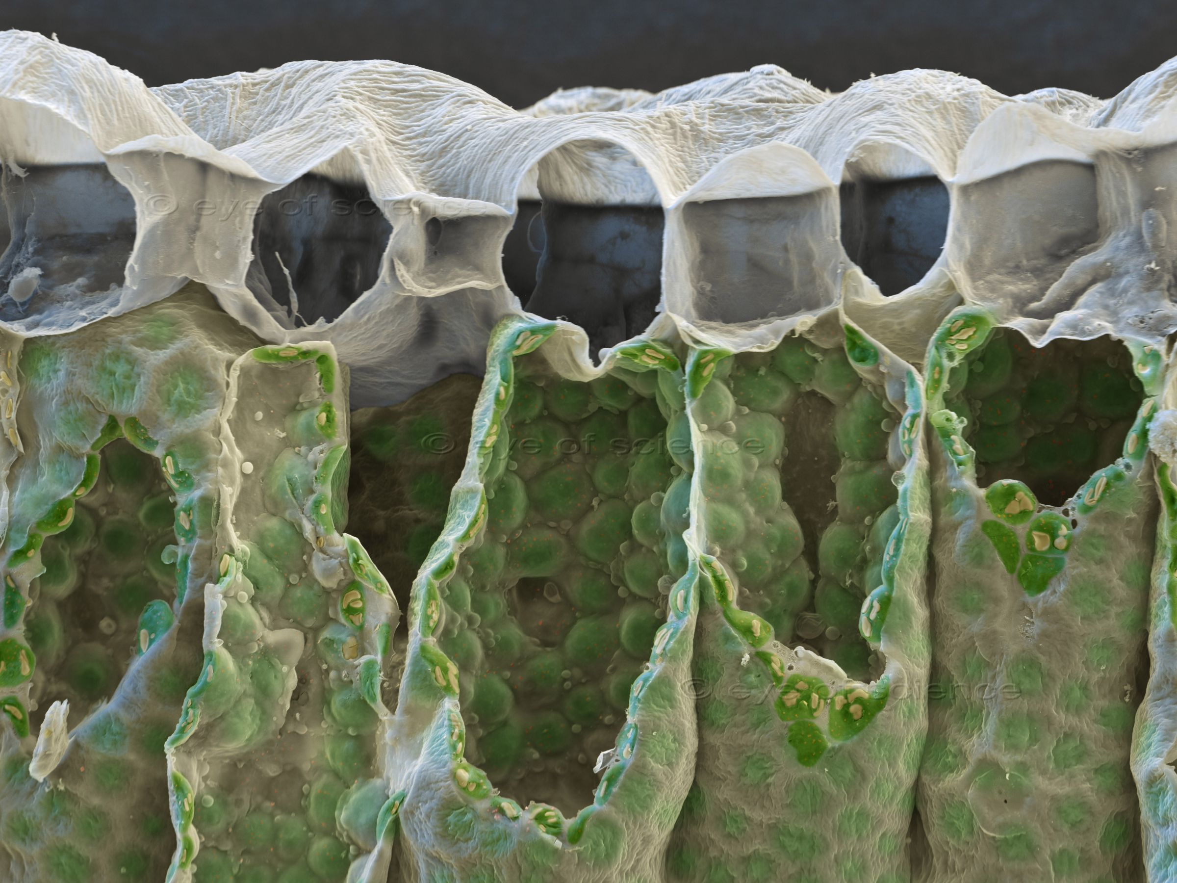
Chloroplasts in a leaf of Solanum nigra. Freeze fraction. Scanning Electron Microscope, 1000:1 (when 15cms wide)
SEM of a Stomata
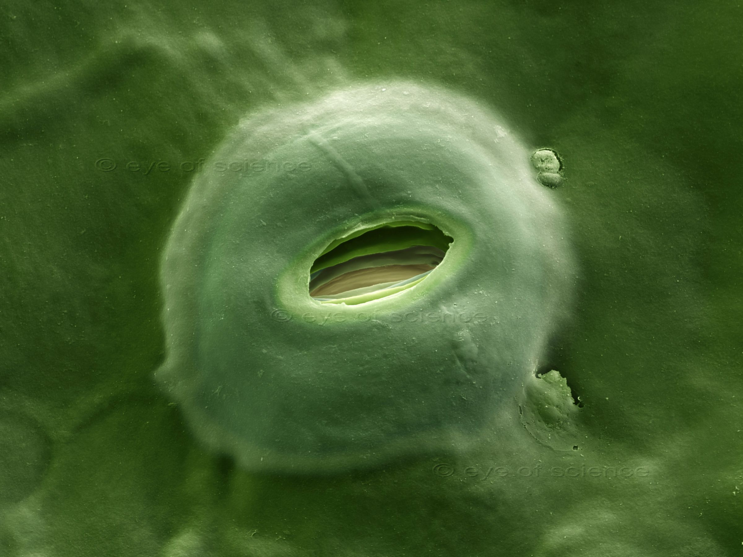
Stomata are the openings for breathing in plant leaves. There the gas exchange CO2 - O2 takes place. Scanning Electron Microscope, 4000:1 (when 15cms wide)
Pollen seen with the Scanning Electron Microscope
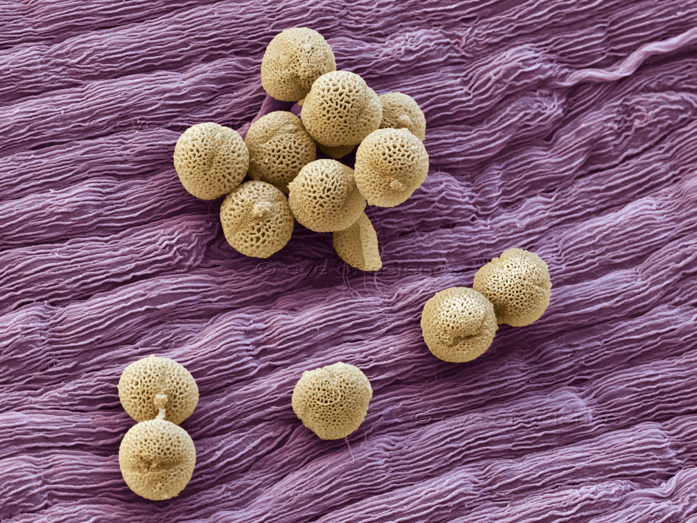
Pollen on a blossom of syringa or lilac. Scanning Electron Microscope, 630:1 (when 15cms wide)
Scanning Electron Micrograph of Chloroplasts
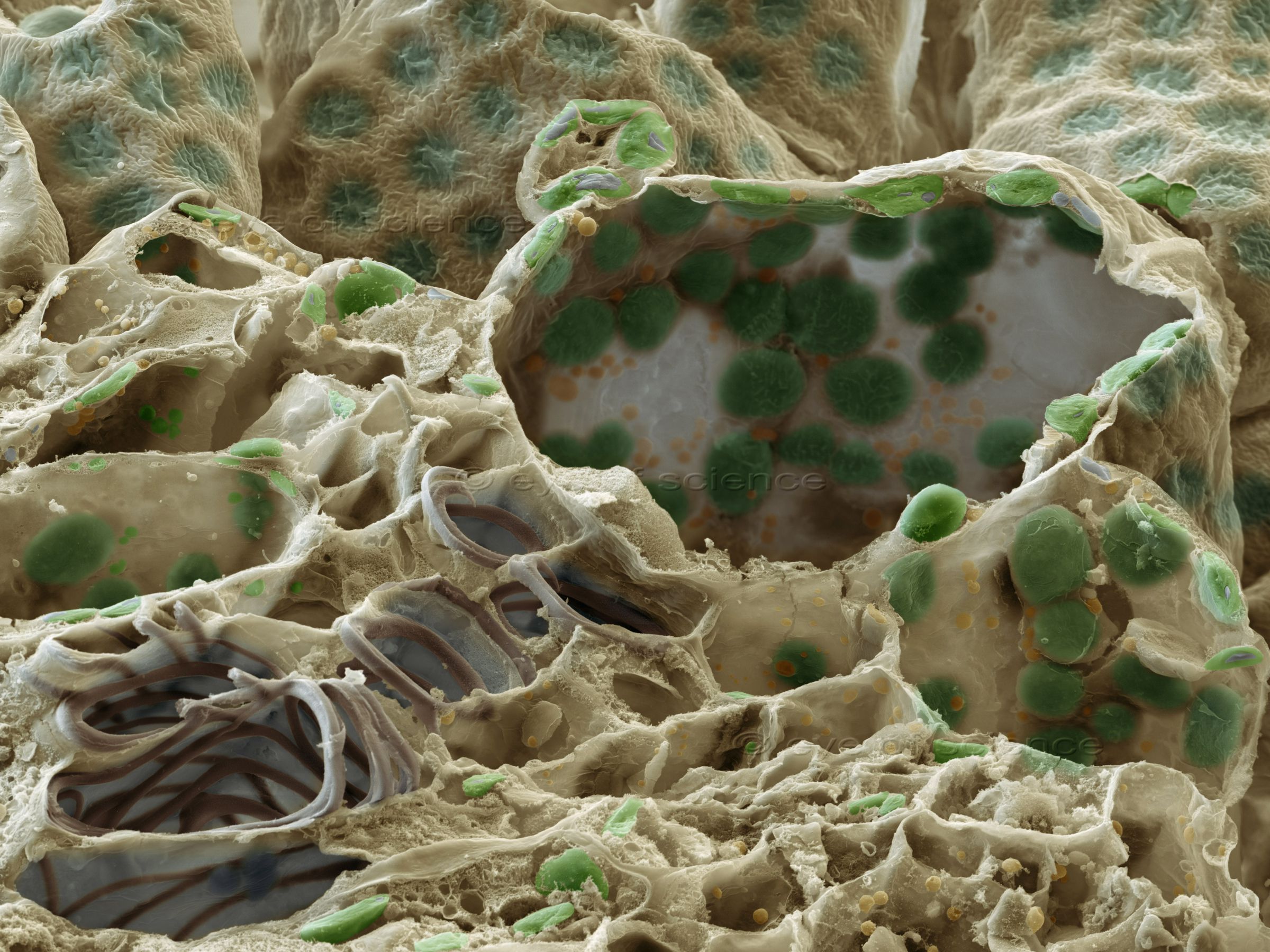
Vascular bundles (lower left) and chloroplasts (green) can be seen in this freeze fraction of a leaf. Scanning Electron Microscope, 1000:1 (when 15cms wide)
SEM image of a Freshwater Alga
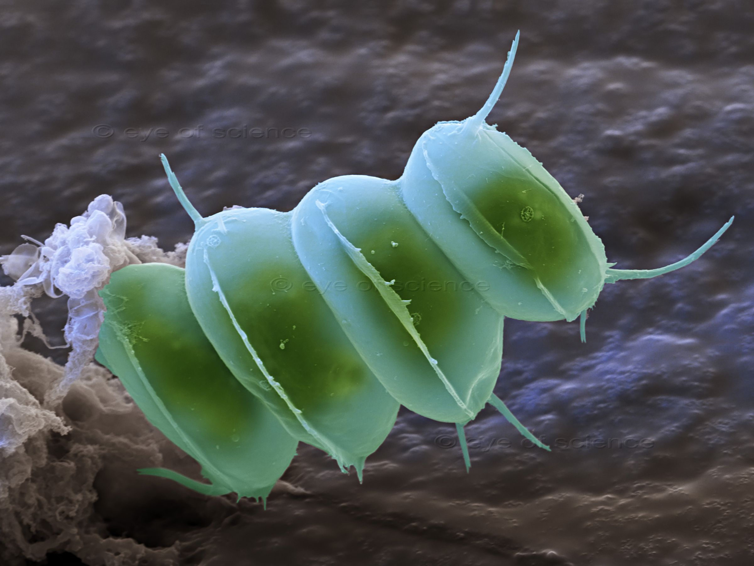
Freshwater alga from a pond. Scanning Electron Microscope, 9000:1 (when 15cms wide



© Copyright for all images on this site by eye of science. All rights reserved.