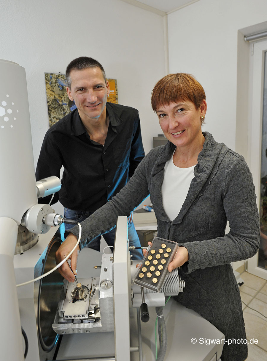
eye of science
founded 1995 by:
Nicole Ottawa
Biologist
1985-1993 Studies of biology at the "Freie Universität Berlin", degree as dipl. biol.
1993-1994 Internship at the Institute for scientific photography, M. Kage
1994 Course in electron microscopy, University of Münster
Oliver Meckes
Photographer
1976 first contacts with microscopy and photography
1983-1985 Vocational training as photographer, starting underwater photography
1986-1989 Internship as a micro photographer and
1989-1994 Scientific photographer at the Institute for scientific photography, M. Kage
STATEMENT
eye of science on artificial intelligence (AI)
eye of science's images are created with the greatest effort and according to scientific criteria in order to depict nature as accurately as possible. For this reason, Eye of Science expressly prohibits the use of its copyrighted images to generate AI content. While AI-generated images may appear realistic, they are the result of algorithms and often lack a real scientific basis. Therefore, they must be clearly labeled as such.
Equipment
.
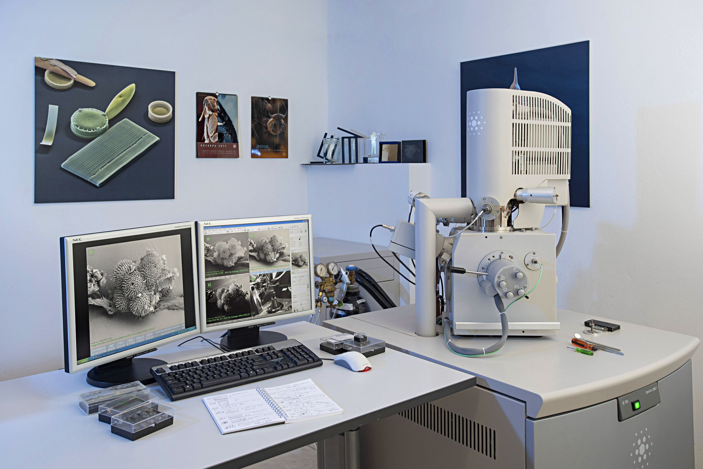
Scanning Electron Microscopes
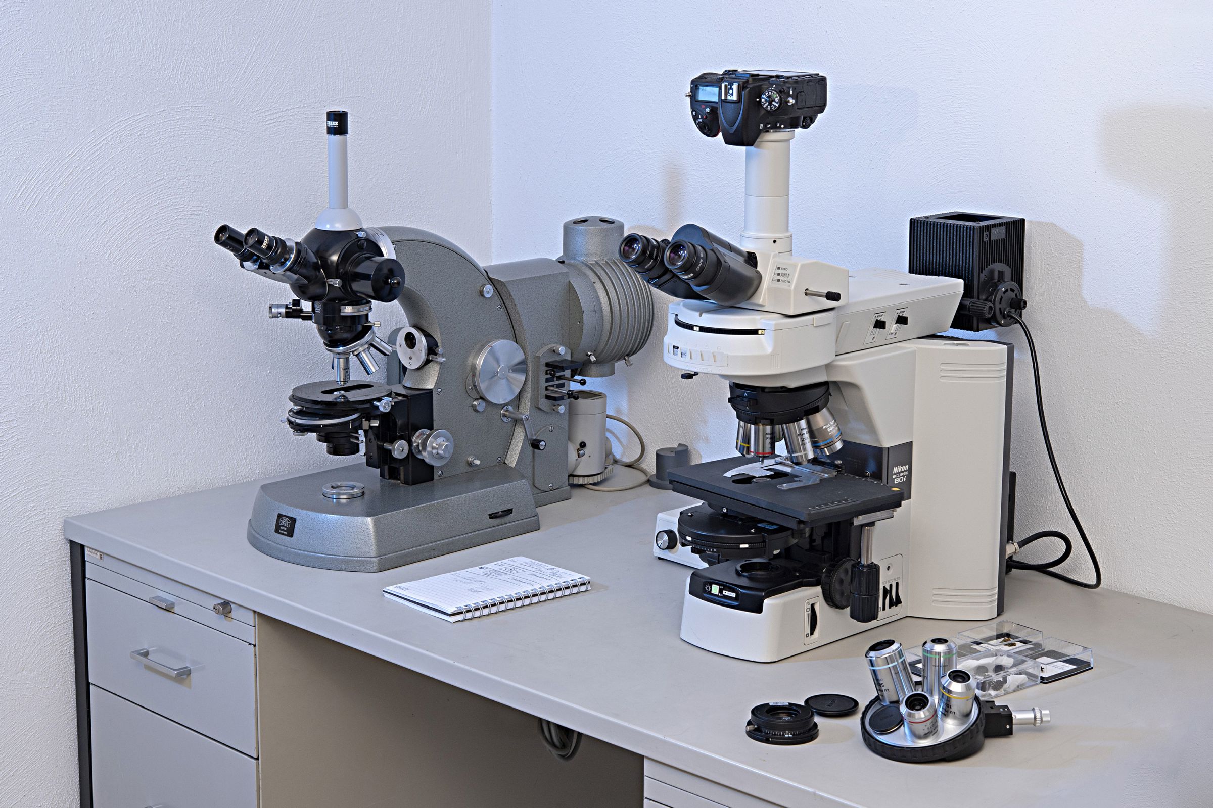
Light Microscopy
Nikon Eclipse 800 laboratory microscope with plan apo-chromatic lenses, polarization, brightfield, darkfield, differential interference contrast and self-made extensions. It can be combined with different camera systems for photography and video.
Zeiss photo microscope with plan- and planapochromatic optics up to 1,000x magnification; rare interference contrast "Jamin Lebedeff".

Specimen Preparation
Stereo microscopes for sample preparation, finest tweezers, needles, hairs to manipulate the specimen. Chemicals for fixation and dehydration. For the required colour imaging of the SEM images several powerful computers are in use.
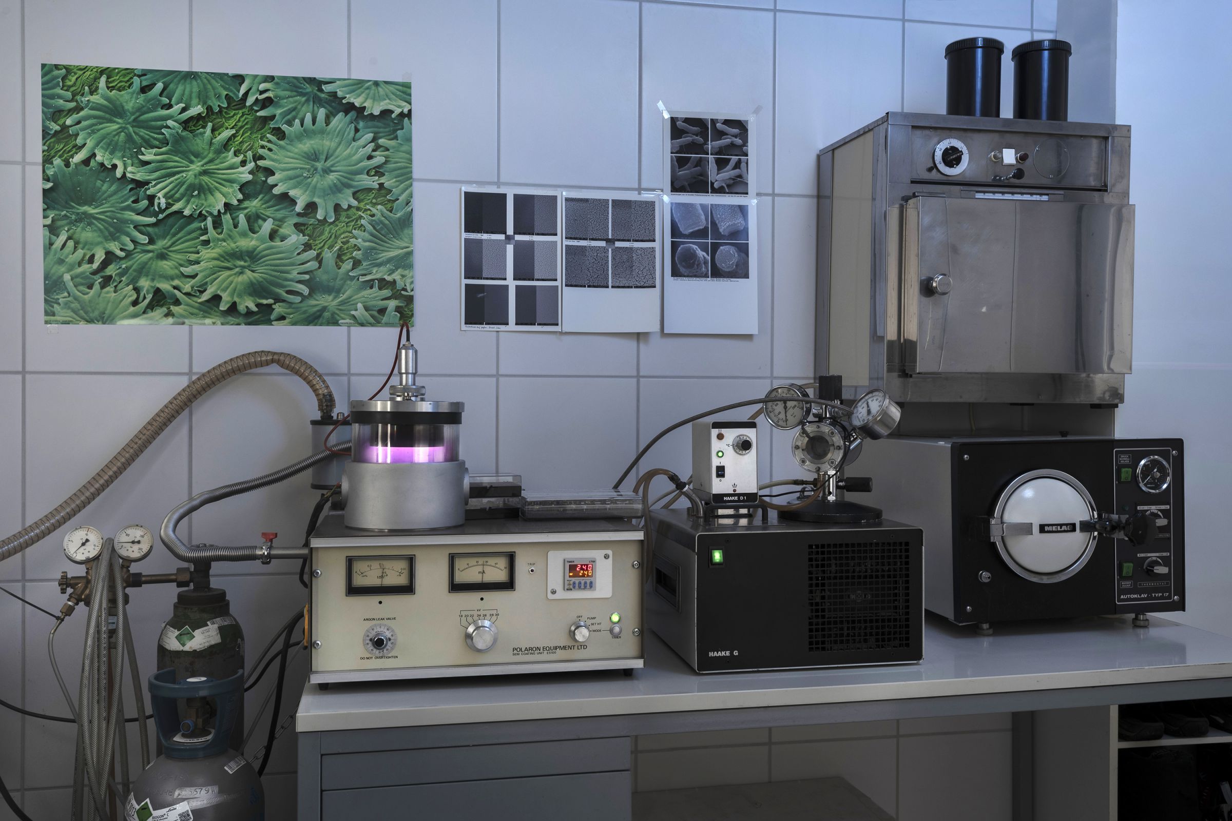
Laboratory
Preparation laboratory for scanning electron microscopy with critical point drying apparatus and sputter-coating with gold or palladium coating. Various equipment for cutting, grinding or etching of technical samples. Incubator for cells and bacteria, autoclave.
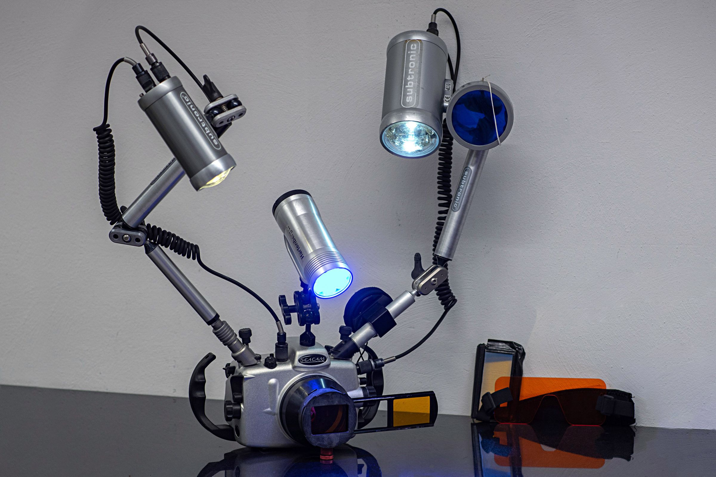
Underwater Photography
Seacam Underwater Housing, equipped with dome and macro-ports, containing Nikon D750 camera with 15mm, 17-35mm, 60mm, 105mm lenses with "Wet Diopter". Several Subtronic flashes and Hartenberger high power LED lamp. Selfmade fluorescent equipment with filters for flash and camera.
© Copyright for all images on this site by eye of science. All rights reserved.