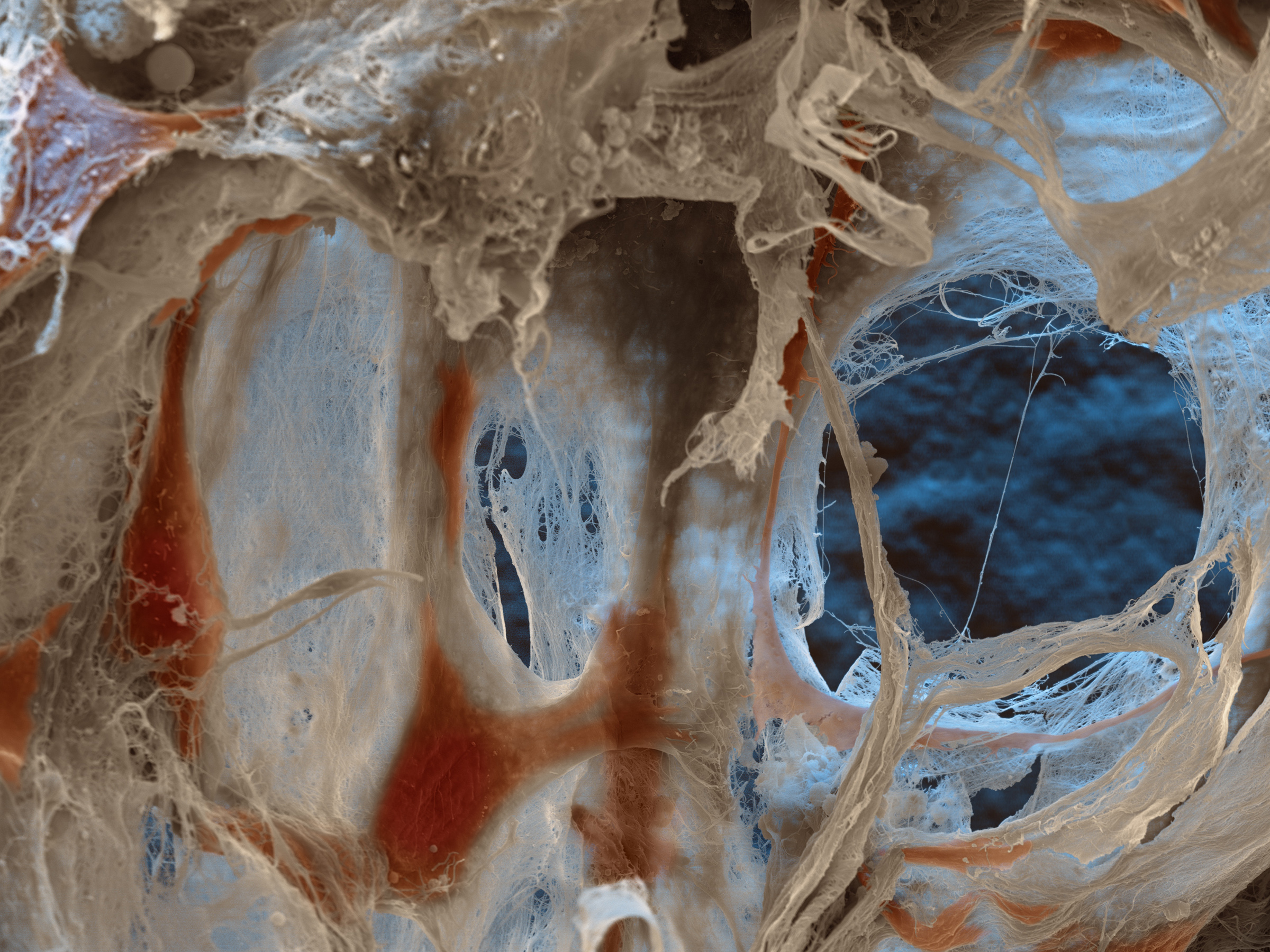
Microphotography for Life-Sciences
The scanning electron microscope is our "camera" for stunning images for life sciences. Our focus is on aesthetics and scientific correctness in equal measure. We do all the preparation for the SEM in our own laboratory. Bacteria, drugs, cell clusters, organoids, yeasts, fungi, blood, organs, culture media? No matter in which area of life science: We will find a way to transform your research into eye-catching images!
Fallopian tube
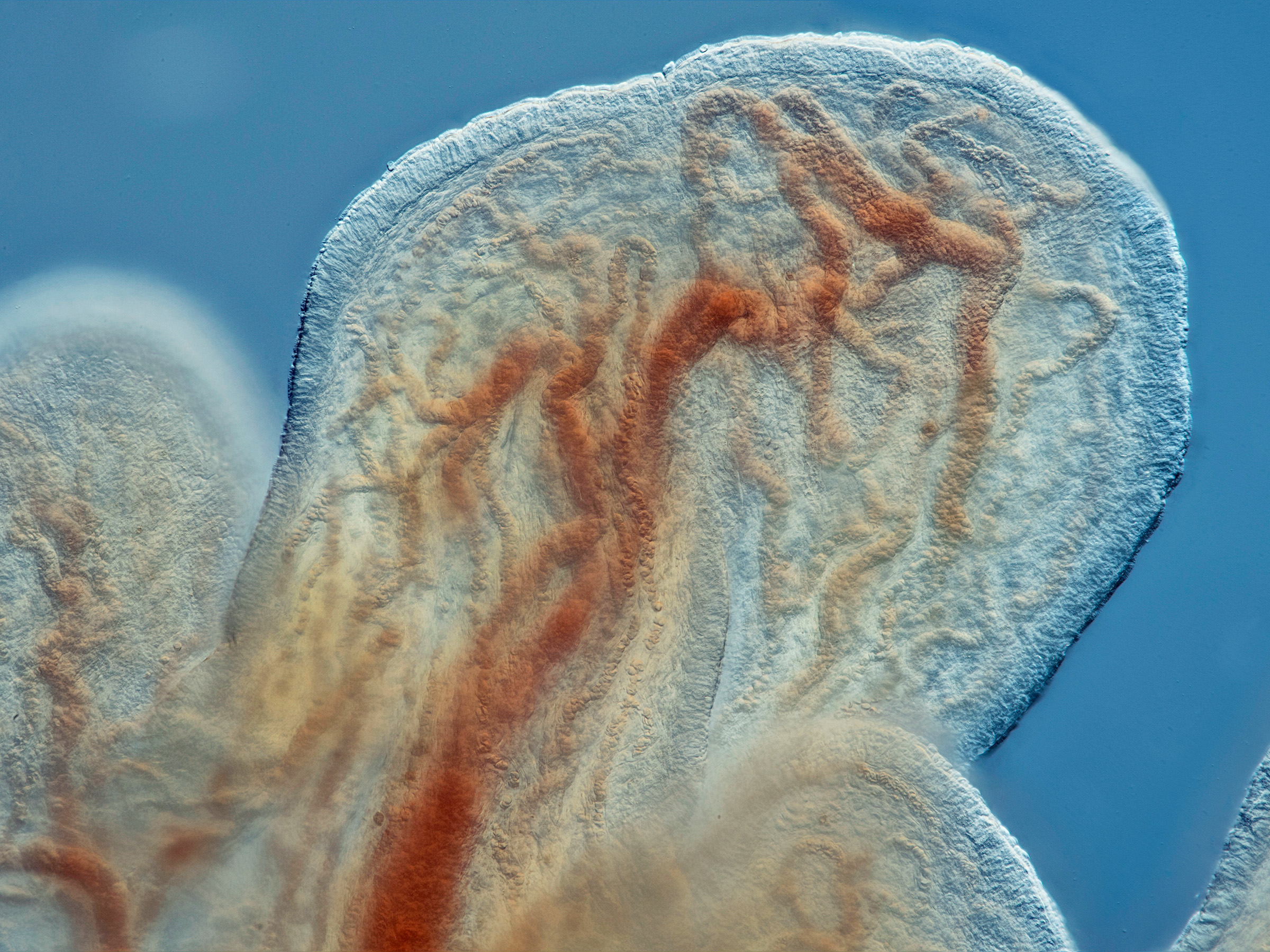
Research into organ function is an important part of "life sciences". This fallopian tube was captured with the light microscope in DIC contrasting. To get more depth of field it was stacked from app. 15 single images. Light microscopic image at 40x magnification.
Cancer
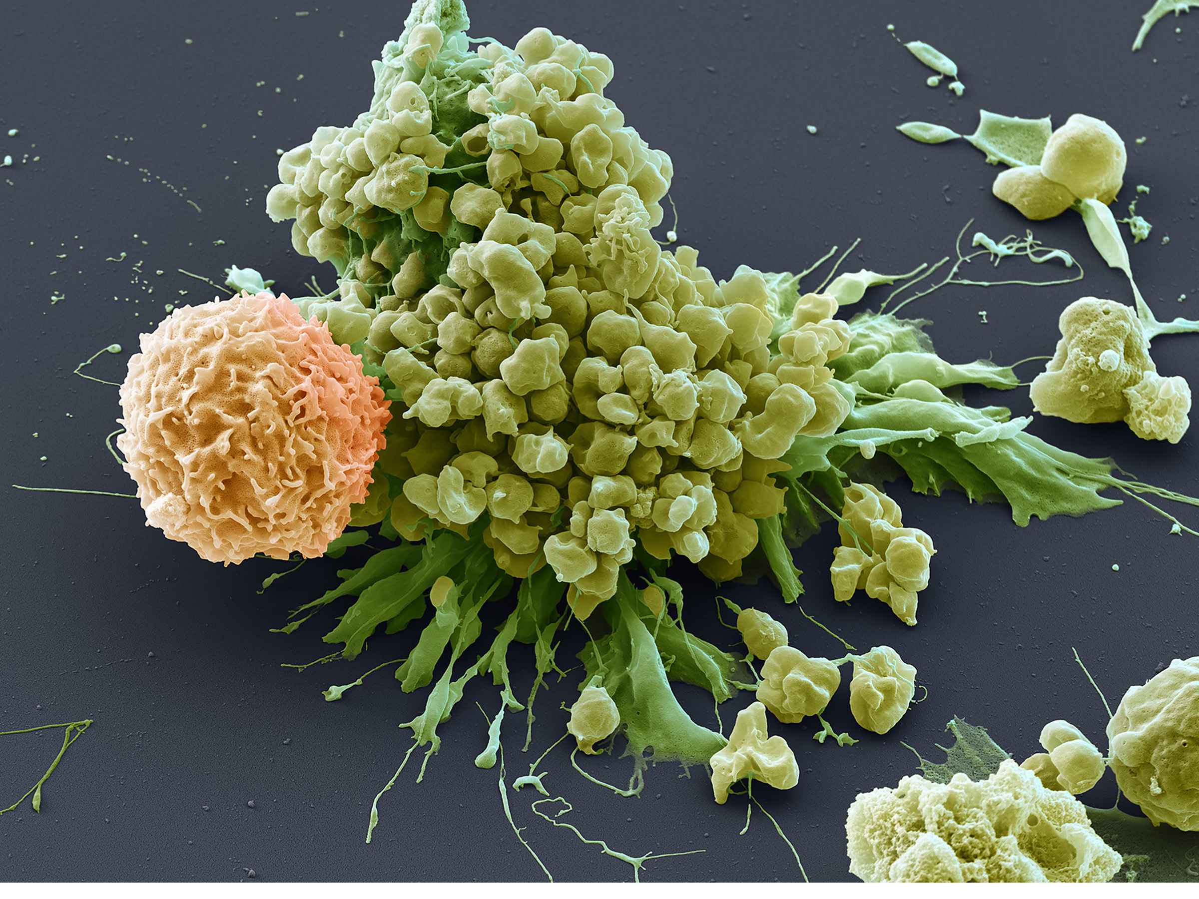
Life science discovered the mechanism of apoptosis and antigen function. This Scanning electron micrograph shows a chimeric antigen receptor cell (CAR) attacking a cancer cell. The cancer cell already started apoptosis. Magnification 5000x (when 15cms wide)
Leishmania
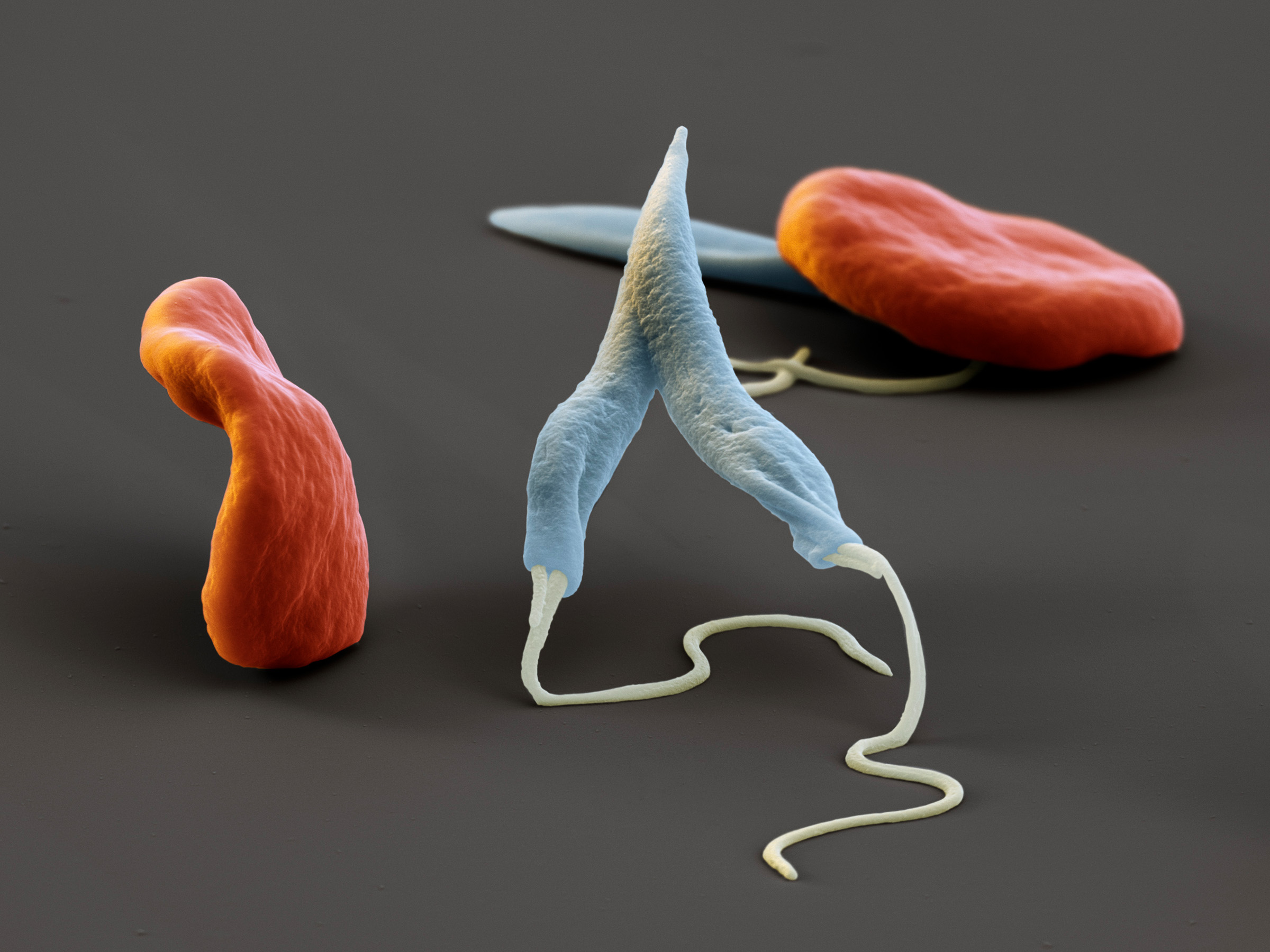
There are still a lot of diseases that have to be observed by life sciences. Leishmania parasites among red blood cells seen with the Scanning Electron Microscope. Leishmaniosis is transmitted by a bite of the sandfly. Magnification: 7000x when 15 cms wide.
Aphidius colemani
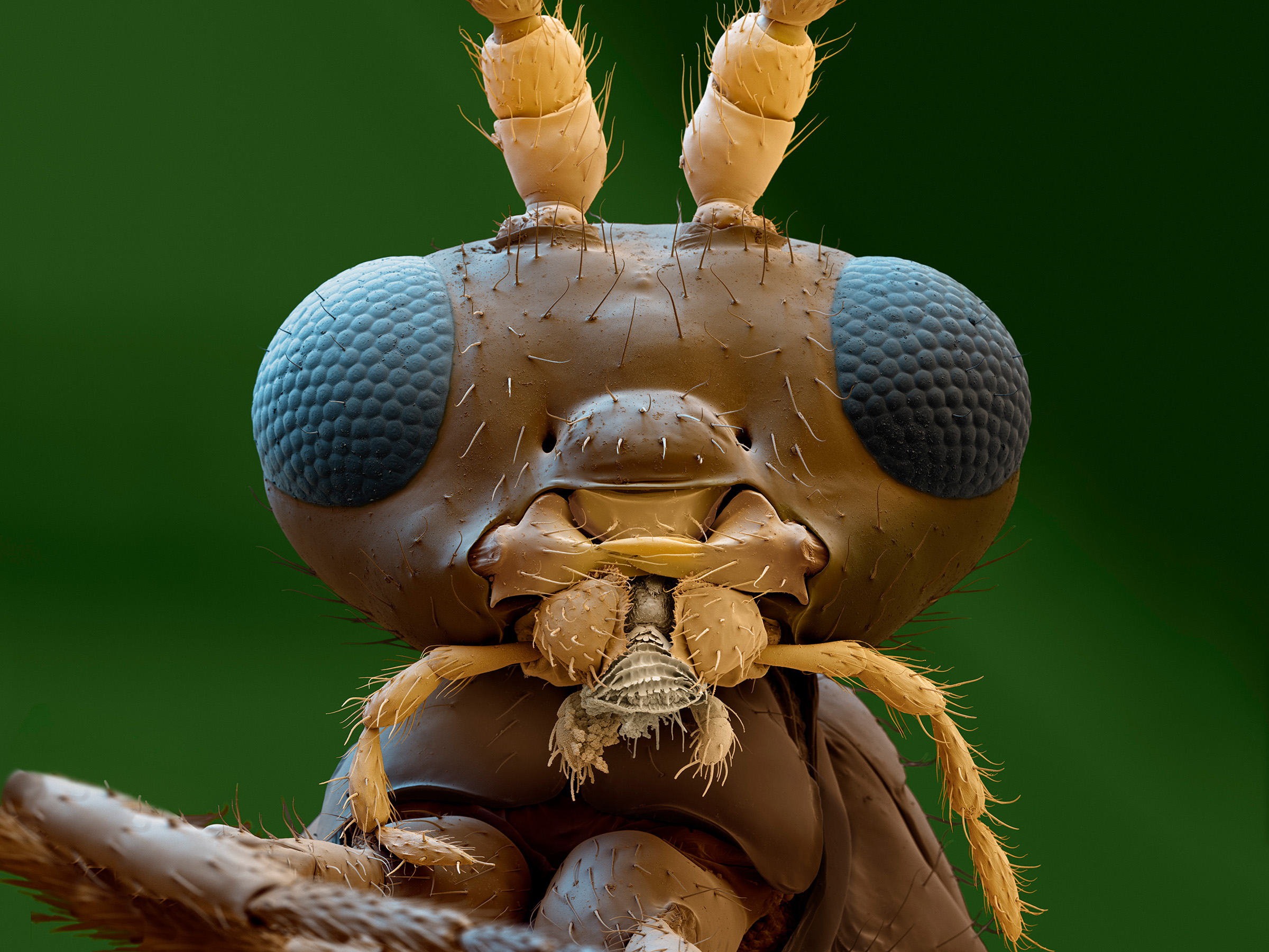
Scanning electron micrograph of Aphidius colemani, a very small parasitic wasp. It is used in crop protection, because their larva feed on aphids. Magnified 200x (when 15cms wide). Life science helps to find solutions to multiply ther population.
For many years we produced micrographs for calendars, advertising or exhibitions showing images from life sciences.
eye of science can look back on almost three decades of experience in preparation, photography and digital image processing in scanning electron microscopy. Over the years the main goal always was life sciences. No matter if bacteria, plants, animals or the human body. We made all kinds of specimen preparation in life sciences for electron microscopy. Aesthetic representation while maintaining scientific authenticity is always in focus. The images are ordered by scientific publishers, chemical and pharmaceutical industries and popular natural science magazines. Via the menu on top you will find examples of our work on electron microscopy, as well as light microscopy and underwater photography, grouped by topic.
© Copyright for all images on this site by eye of science. All rights reserved.