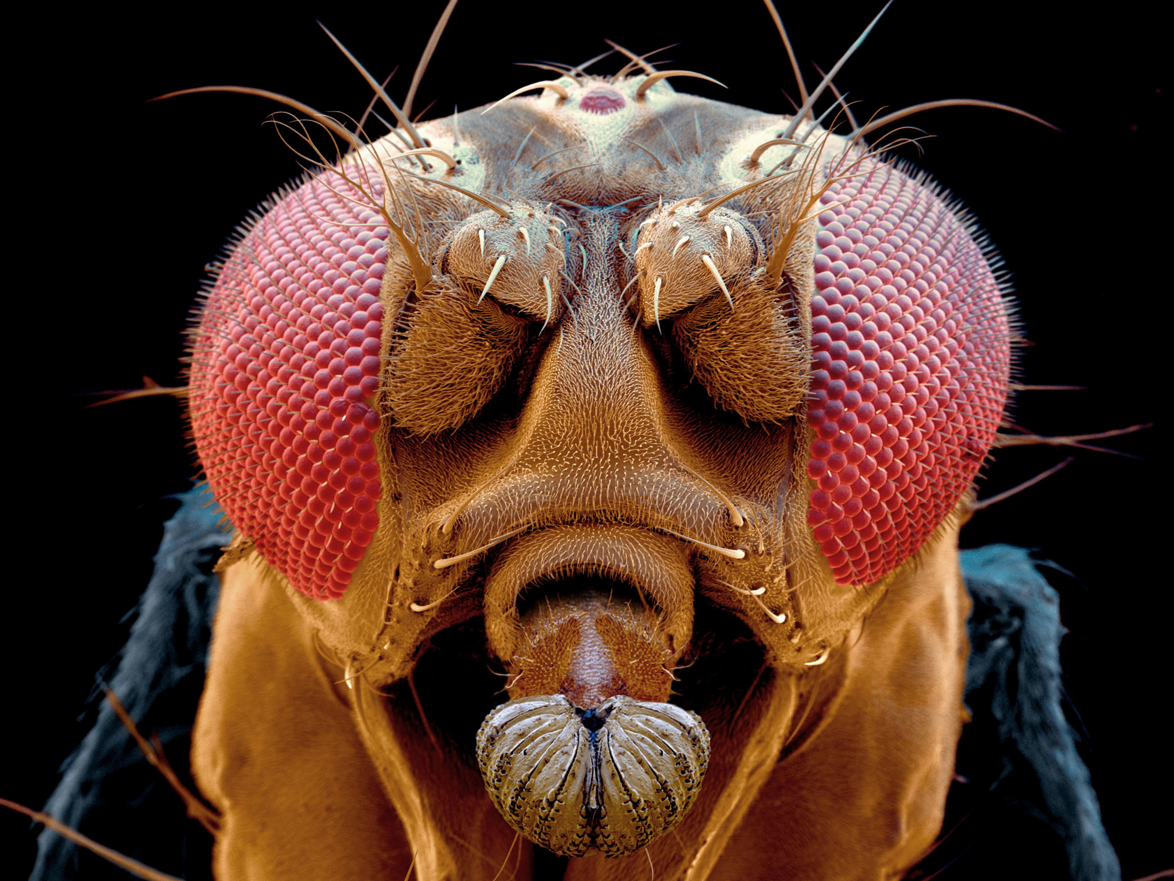
Microphotography supports comprehension
Education, especially in the field of natural sciences, benefits from microscopic images in a wide range of fields. Our "portrait" of the fruit fly has become an icon in school textbooks, but we offer much more. Light microscopic images of organs, plant sections, blood smears are an integral part of biological and medical education. Electron microscopy depicts the cell structure, the relationships between cells or is able to spatially perfectly image entire (micro-) organisms or organs and thus transport knowledge for education.
Pollination
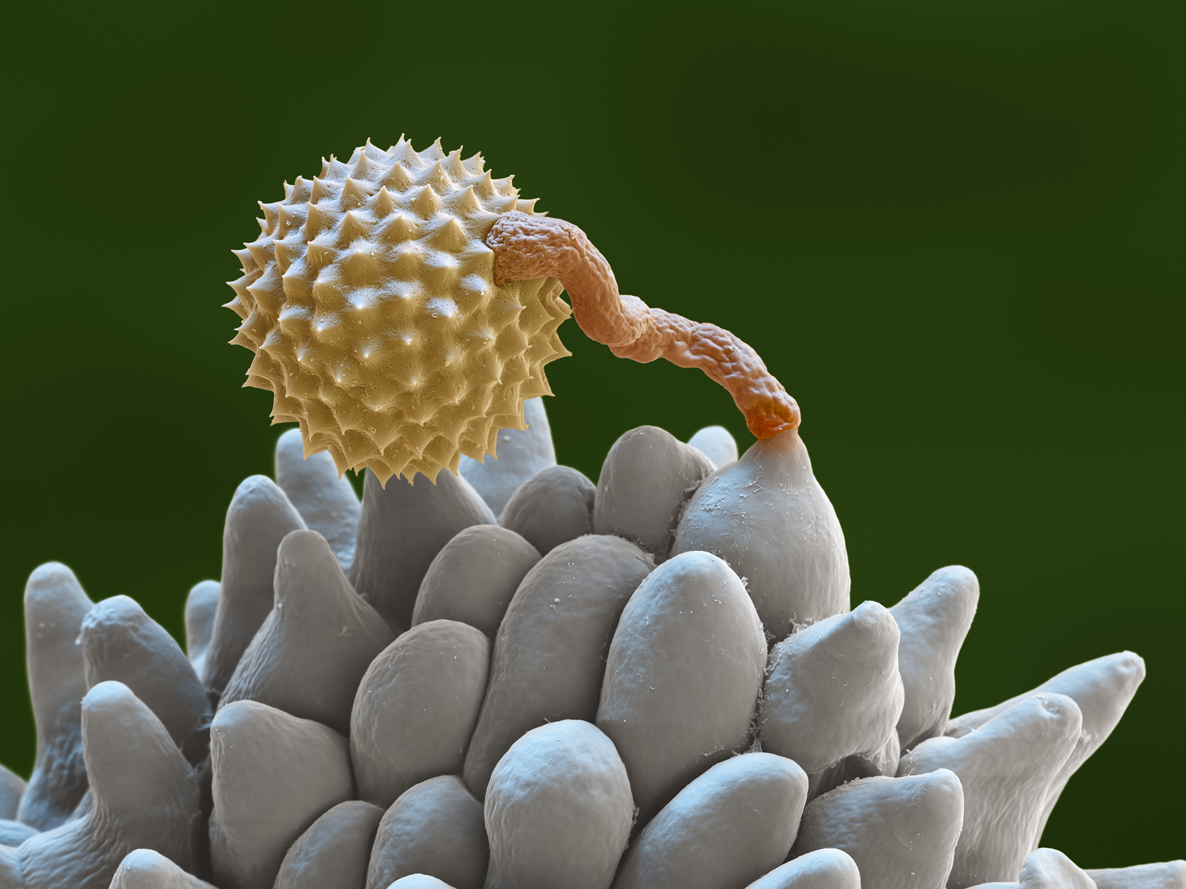
A germinating pollen grain on a stigma captured with the scanning electron microscope may teach the function of fertilization.
Chromosomes

A light micrograph showing different stages of mitosis in Allium cepa (onion root) is found in most biology school books in the higher classes.
Escherichia coli
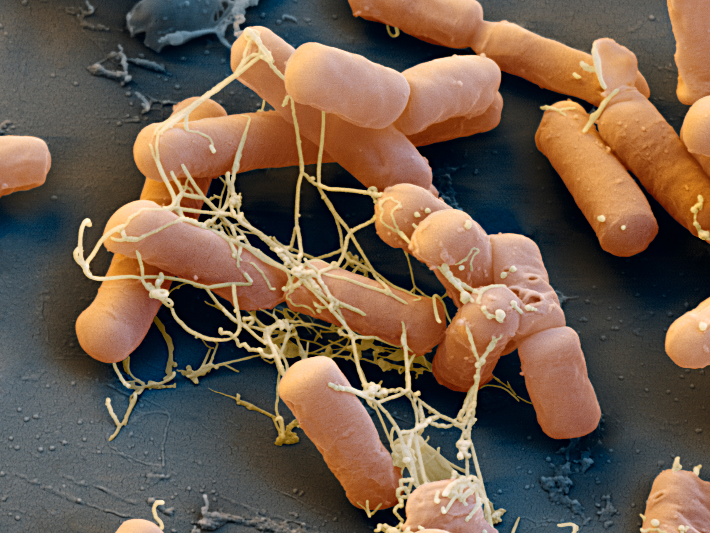
One of the standard bacteria in research: Escherichia coli, seen with the scanning electron microscope. One of the "must" organisms in biological education.
Bacteriophage
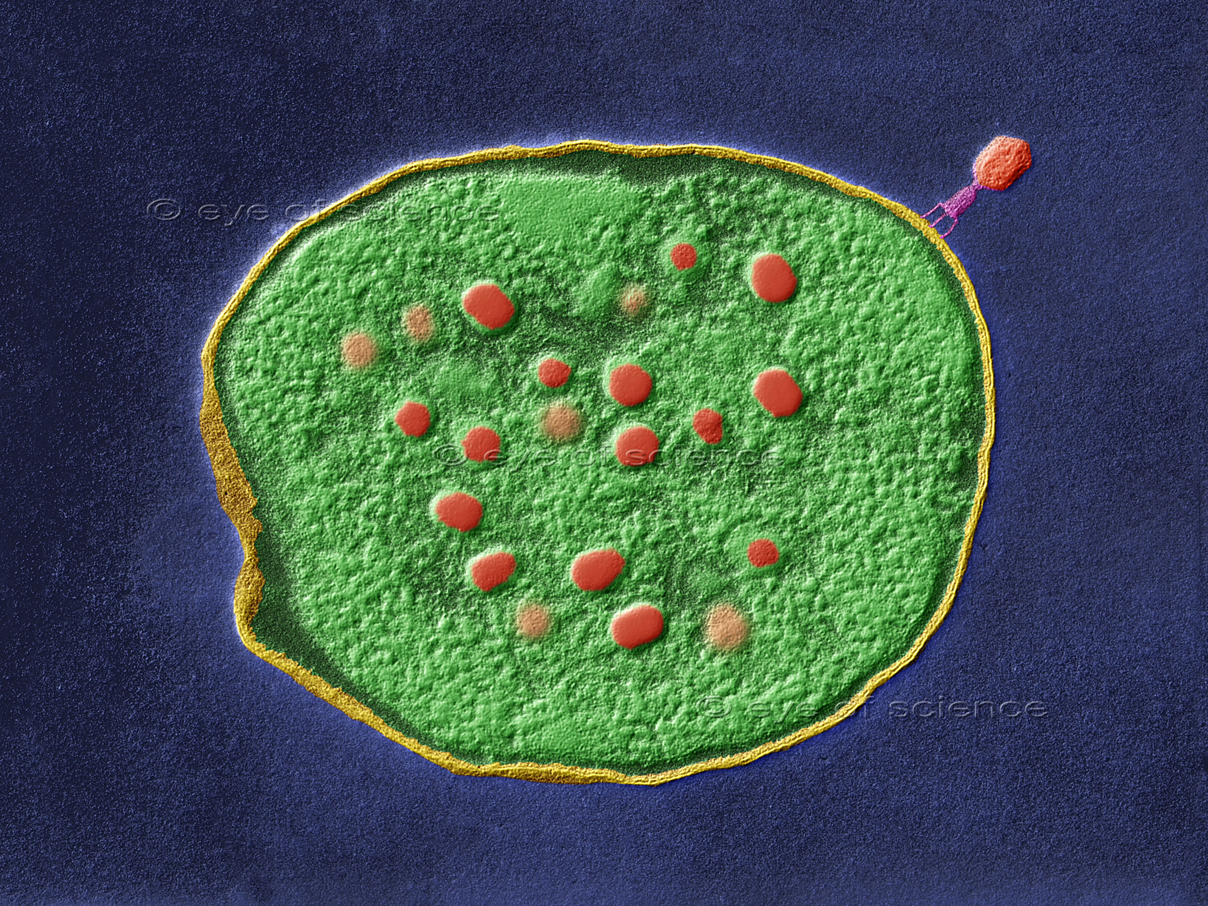
TEM image of a T phage on Escherichia coli. A single bacterium containing assembling phages (red dots): Learning how infection works.
Micrographs support visual understanding and allow fascinated learning
eye of science has many years of experience in preparation, photography and digital imaging in scanning electron microscopy. We have been working with educational publishers from the start. Our micrographs are designed to help teachers bring visual understanding into their lessons. Colored SEM images are a great help for understanding biological systems and relationships. Because we prepare and photograph all samples in our own laboratory, we can guarantee authentic and realistic use for educational purposes. Via the menu on top you will find examples of our work on electron microscopy, as well as light microscopy and underwater photography, grouped by topic.
© Copyright for all images on this site by eye of science. All rights reserved.